vesicle_triangulator — Example output : ch1
Per-channel intermediates for channel 01 of the example run. These files are the upstream products that feed the joint triangulation in ../mixed/; they are kept next to the final outputs so you can inspect how the ch1 vesicle points were detected before being triangulated against ch2.
- 📄 ch1_components.png – Download
- 📄 ch1_overlay.png – Download
- 📄 ch1_points.csv – Download
- 📄 ch1_thresh.png – Download
📜 Files in this folder
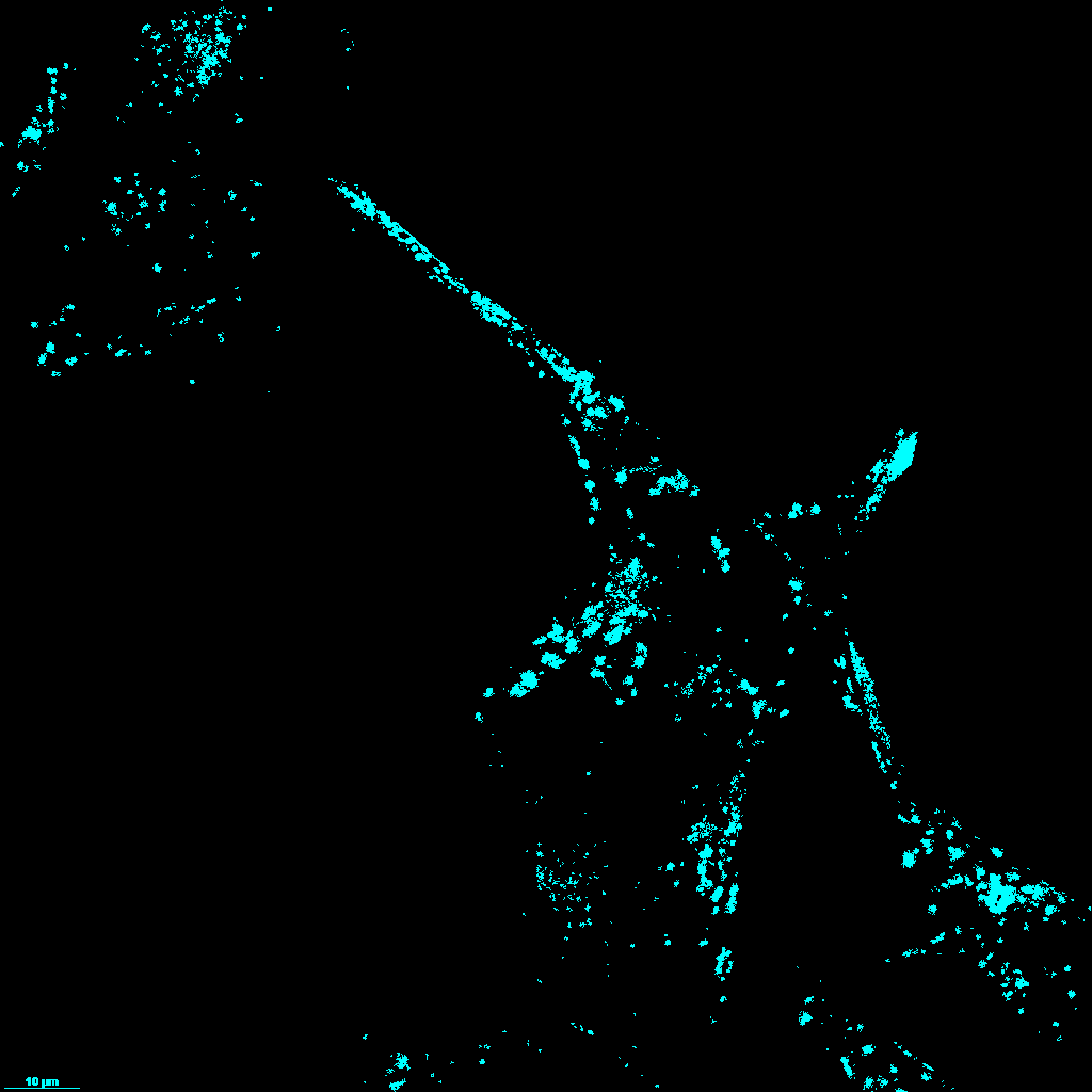
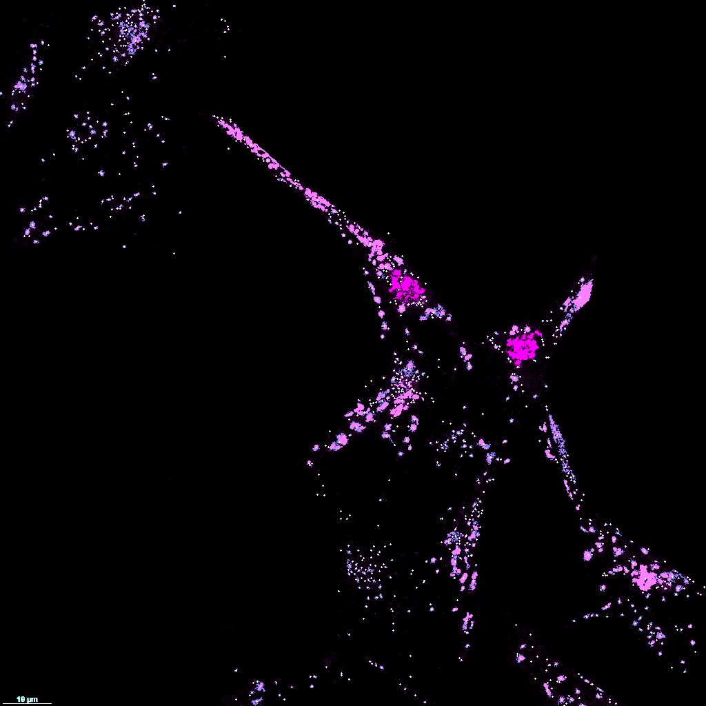
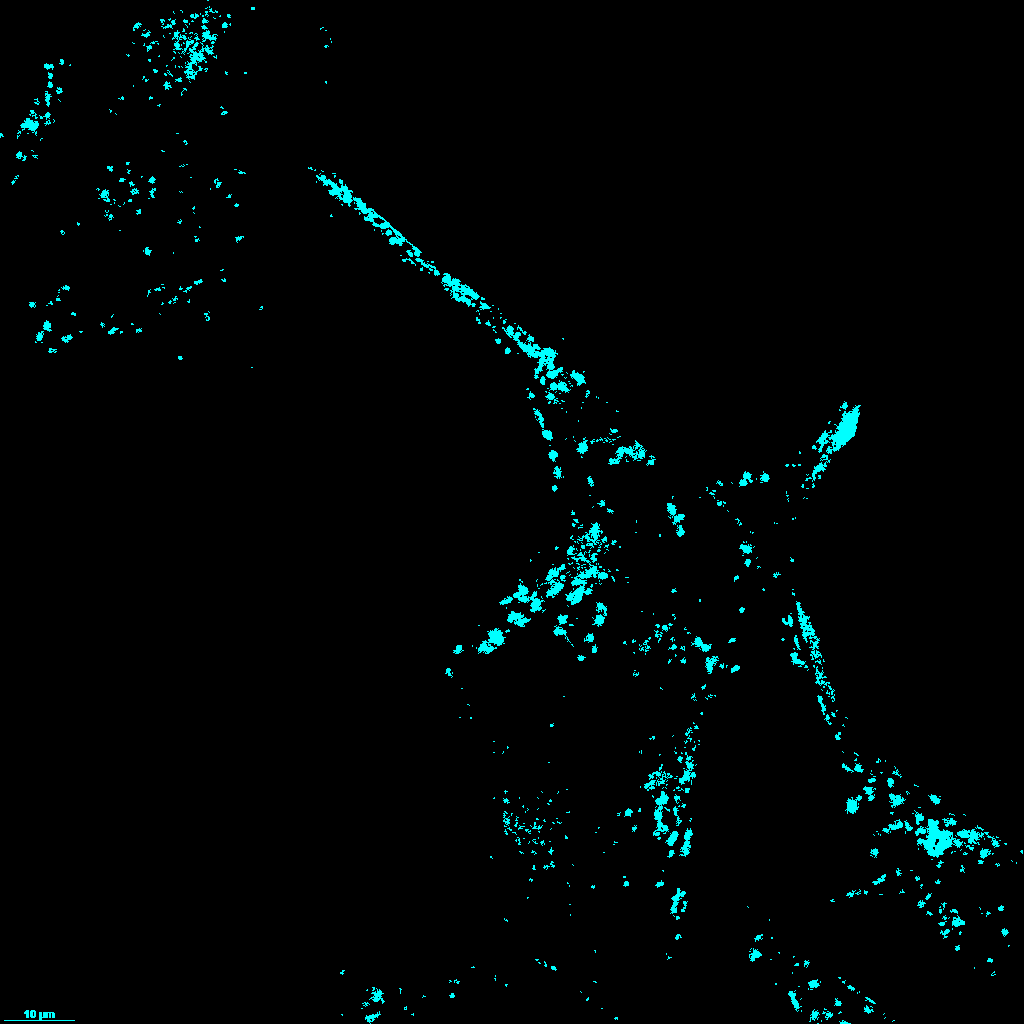
ch1_thresh.png— Binary threshold mask for channel 01: the raw pixel-level result of the ch1 threshold setting. This is what you see in the GUI right after clicking “Threshold Ch1”.ch1_components.png— Connected-component labeling ofch1_thresh.png: each distinct vesicle is colorized with its own component ID so you can spot-check that neighboring vesicles were correctly split (or not merged).ch1_points.csv— Point centroids extracted from the components as(x, y)coordinates, one row per vesicle. This is the input to the triangulator — all ch1 vertices in the mesh come from here.ch1_overlay.png— Centroids fromch1_points.csvdrawn on top of the original microscopy image, so you can verify visually that every detected point lands on a real vesicle.